


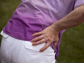
The hip joint (HJ) is a complex joint made up of several bones: the femur, pubis, ilium, and ischium. It is surrounded by bags around the joints and a corset of strong muscular ligaments and protected by subcutaneous fat and skin.
The ilium, ischium, and pubis form the pelvis and are connected by the hyaline cartilage at the acetabulum. These bones fuse together before the age of 16.
A distinctive feature of the femoral joint is the structure of the acetabulum, the upper and lateral sides of which are only partially covered with cartilage. The middle and lower segments are occupied by adipose tissue and the femoral ligament, encased in the synovium.
reason
Hip pain can cause damage to components within the joint or nearby structures:
- skin and subcutaneous tissue;
- muscles and ligaments;
- synovial bag;
- The acetabular labrum (the cartilaginous edge that extends along the rim of the acetabulum);
- The articular surfaces of the femur or pelvis.
Pain in the joint area is caused by inflammation or damage to the integrity of its constituent structures. Most often, pain occurs when infection enters the joint cavity (septic arthritis) and autoimmune lesions (rheumatoid arthritis and reactive arthritis).
Also common are mechanical injuries, which result in damage to the epiphysis of bones, ligaments, synovium, and other tissues. Trauma is more likely to occur when active people and athletes experience high-intensity physical exertion.
Also at risk are older adults with pelvic pain due to degenerative dystrophic changes in the cartilage, and children and adolescents during hormonal changes.
Left or right hip pain is caused by metabolic diseases such as diabetes, pseudogout, and obesity.
The full list of possible diseases is as follows:
- Perth disease;
- joint;
- Koenig's disease;
- Diabetic joint disease;
- pseudogout;
- Intermittent hydrojoint (intermittent edema of the joints);
- rickets;
- Reactive, rheumatoid and infectious arthritis;
- juvenile epiphyseal osteolysis;
- Hurt.
Perth disease
Perthes disease disrupts the blood supply to the femoral head, causing sterile necrosis (death) of cartilage tissue. Most children (mostly boys) under the age of 14 suffer.
The main symptom of Perthes disease is persistent pain in the hip joint that worsens with walking. Many times, children complain of pain in their legs from their hips and begin to limp.
In the initial stage, symptoms are mild, leading to a late diagnosis when an impression (intra-articular) fracture has already occurred. The destructive process is accompanied by increased pain, swelling of soft tissues and stiffness of limb movements. The patient cannot turn, rotate, bend, or straighten the thigh outward. It is also difficult to move the leg to the side.
Violations of the autonomic nervous system were also observed: feet became cold and pale, with profuse sweating. Sometimes the body temperature rises to sub-calorific values.
Reference: Lesions in Perthes disease can be unilateral or bilateral. In most cases, one of the joints is less affected and recovers faster.
joint
Osteoarthritis of the hip, called hip disease, is primarily diagnosed in older adults. The disease progresses slowly but causes irreversible changes. The pathological process begins with damage to the cartilage, which becomes thinner due to the increased density and viscosity of the synovial fluid.
The development of hip arthropathy results in joint deformity, muscle atrophy, and significant restriction of movement up to complete immobility. Arthropathy pain syndrome has a wavy (non-permanent) feature that is located on the outside of the thigh but can spread to the groin, buttocks, and lower back.
In the second stage of arthropathy, pain covers the inner thigh and sometimes extends to the knee. Hip pain increases as the disease progresses and sometimes subsides with rest.
Hip disease is primary and secondary. Primary hip arthropathy develops in the context of osteochondrosis or knee arthropathy. Prerequisites for secondary hip arthropathy may be hip dysplasia, congenital hip dislocation, Perthes disease, arthritis and trauma (dislocations and fractures).
Koenig's disease
If the thigh is injured on one side of the joint area, the cause may be death (necrosis) of cartilage tissue - Koenig's disease. The disease most commonly occurs in young men aged 16-30 who complain of pain, reduced range of motion and periodic "stuck" in the legs.
Koenig disease develops in several stages: first, the cartilage softens, then thickens and begins to separate from the articular surface of the bone. In the third or fourth stage, the necrotic area is rejected and enters the joint cavity. This is due to the accumulation of effusion (fluid), stiffness of movement, and blockage of the left or right joint.
Reference: The presence of "joint mice" in the hip joint leads to the development of hip arthropathy.
Diabetic Arthropathy
Osteoarthropathy, or Charcot's joint, is seen in diabetes, characterized by progressive deformity with pain of varying intensity. Due to the pathological changes of nerve fibers, the sensitivity of this disease is drastically reduced, so that the expression of pain sensation is rather weak or completely absent.
Diabetic arthropathy occurs with a long course of diabetes and is one of its complications. It occurs most often in women who have not received adequate or ineffective treatment. It should be noted that the hip joint is rarely affected.
pseudogout
As calcium metabolism is disrupted, calcium crystals begin to build up in joint tissue and develop chondrocalcinosis or pseudogout. The etiology and symptoms are named for their resemblance to gout, which is characterized by a paroxysmal course.
Sudden, severe, severe pain: The affected area becomes red and swollen, and is hot to the touch. Inflammatory flare-ups last from hours to weeks, and then everything passes. With chondrocalcinosis, pain may occur on the left or right side of the pelvis.
In the vast majority of cases, pseudogout occurs for no apparent reason, and calcium disturbances cannot be detected even during examination. It is speculated that the cause of the disease is a local metabolic disturbance within the joints. In one percent of patients, chondrocalcinosis develops in the context of an existing systemic disease (diabetes, renal failure, hemochromatosis, hypothyroidism, etc. ).
synovial chondromatosis
Arthrochondromatosis, or synovial cartilage islet metaplasia, primarily affects large joints, including the hip. Mostly, this pathology occurs in middle-aged and older men, but there are also cases of congenital chondromatosis.
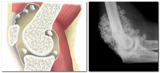
In chondromatosis, the synovium degenerates into cartilage or bony tissue, resulting in the formation of cartilage or bony bodies up to 5 cm in size in the joint cavity.
The clinical presentation of insular metaplasia is similar to that of arthritis: the patient is concerned about hip pain, limited leg movement, and a characteristic creaking noise is heard during movement.
Since chondromatosis is a dysplastic process with the formation of cartilage bodies, the occurrence of "joint mice" cannot be ruled out. In this case, the "mouse" may get stuck between the articular surfaces of the bones, which will result in a partial or complete blockage of the joint. The joint remains blocked until the cartilaginous body enters the capsule, after which full motion can be resumed.
Reference: Frequent or prolonged joint jamming can trigger the development of hip arthropathy. Complications of synovial chondromatosis are stiffness (contractures) and muscle wasting.
arthritis
Arthritis is an inflammation of the joint surface of the acetabulum and femur. The failure of the hip joint is called hip arthritis, with dull pain in the back of the thigh and groin.
There are various types of arthritis, the most common being the hip joint affected by its infected form. Other species are diagnosed much less frequently. Why does septic arthritis occur? After the entry of bacteria and viruses into the joint cavity, the development of pathology begins.
The clinical presentation of septic arthritis can vary depending on the type of microorganism that causes it. However, 5 characteristic signs were observed in all patients:
- right or left leg joint pain syndrome (also bilateral lesions);
- swollen and swollen joints;
- redness of the skin;
- decreased exercise capacity;
- Elevated body temperature.
At the onset of the disease, patients experience severe pain, especially when getting up from a sitting position. The joints are almost always sore because the pain cannot stand or sit. It should be noted that the infectious form of arthritis is always accompanied by fever, chills, headache, weakness and nausea.
juvenile epiphysis
The term condylar release literally means the dismantling, destruction of the articular surface of a bone, or rather, the cartilage that covers it. A distinguishing feature of this injury is the cessation of growth in bone length, which results in asymmetry in the lower extremity.
In adults, fractures of the epiphysis occur in which the epiphysis is displaced or ruptured. Destruction of the growth zone epiphysis is only possible during adolescence, so this disease is called juvenile disease.
Juvenile epiphysiolysis is an endocrine-orthopedic pathology based on an imbalance between growth and sex hormones. It is these two groups of hormones that are critical to the normal function of cartilage tissue.
The dominance of growth hormone over sex hormones results in decreased mechanical strength in the growth zone of the femur and displacement of the epiphysis. The end portion of the bone is below and behind the acetabulum.
Typical symptoms of epiphyseal osteolysis are pain on the right or left side of the thigh (depending on the joint affected), lameness, and an unnatural position of the leg. The diseased leg is everted, and the muscles of the buttocks, thighs and calves are atrophied.
treat
To treat Perthes disease, chondroprotective agents are needed to promote cartilage regeneration, and vasoprotective agents are necessary to improve blood circulation. Combination therapy also includes massage, exercise therapy, physiotherapy - UHF, calcium and phosphorus electrophoresis, mud and ground stone applications.
Patients with Perthes disease are advised to remove their limbs and use orthopedic devices (plaster casts) and special beds to prevent deformity of the femoral head.
What to do and what medicine to drink for joint disease depends on the stage of the disease. The following remedies can help relieve pain and slow down the pathological process in stages 1-2:
- nonsteroidal anti-inflammatory drugs (NSAIDs);
- vasodilators;
- Muscle relaxants relax muscles;
- chondroprotective agent;
- hormones (with severe pain);
- Ointments and compresses with anti-inflammatory or chondroprotective properties.
In stages 3-4, patients undergo surgery.
Koenig disease is only treated surgically, during arthroscopic surgery, the affected area of cartilage is removed.
Treatment of diabetic arthropathy involves correcting the underlying disease - diabetes, wearing special stress relief bandages and taking medication. All patients, regardless of disease stage, need to take anti-absorptive drugs - bisphosphonates, as well as drugs containing vitamin D and calcium. To reduce pain and inflammation, NSAIDs and corticosteroids are prescribed. Antibiotics are administered if there are infectious complications.
There is no specific treatment for pseudogout; anti-inflammatory drugs are used to exacerbate. A large accumulation of fluid in the joint is an indication for an intra-articular puncture, during which fluid is pumped and corticosteroid medication is administered.
Hip chondromatosis requires mandatory surgical intervention, and its volume depends on the extent of the lesion. For a few cartilaginous bodies, they are removed by partial synovectomy (removal of the synovium) or minimally invasive arthroscopy (through three punctures). Surgical treatment of progressive chondromatosis can only be radical and can be performed using open arthrotomy or complete (total) synovectomy.
The treatment of acute infectious arthritis consists of the mandatory application of a cast in the hip area, the administration of various medications (NSAIDs, antibiotics, steroids). As the purulent process develops, a series of therapeutic punctures are performed to disinfect the joint.
Treatment of juvenile epiphyseal osteolysis is surgery only. During surgery, a closed repositioning of the bone is performed, for which bone traction is used. The combined part of the bone is then secured with a needle and graft.
Absolutely all hip disorders are serious conditions requiring mandatory medical supervision. Any injury following a fall or impact, along with severe pain, limited mobility, and changes in joint structure, requires emergency medical attention. If there is no trauma, and joints often experience pain of varying intensity, it is necessary to make an appointment with a GP or rheumatologist and be examined.