Arthropathy of shoulder joint

Shoulder ArthropathyShoulder arthritis is a dystrophic lesion of the cartilage plate covering the articular surface of the joint, with subsequent involvement of the underlying bone.

About disease

This disease affects not only the cartilage layer and subchondral bone. The pathological process also gradually involves the joint capsule and ligamentous apparatus, synovium, tendon chambers, and subacromial area.Arthropathy of the shoulder joint at a certain stage can lead to the development of osteoarthritis. This condition is characterized by the following symptoms: chronic pain, reduced joint range of motion, and creaking within the joint when rotating. Most often, people over the age of 40 experience this transition.The main symptoms of shoulder joint are pain and limited arm movement. To verify the diagnosis, imaging methods provide a wealth of information - ultrasound and X-ray scans, computed tomography and magnetic resonance imaging.According to clinical recommendations, conservative treatment is used in the early stage of the disease. In the later stage, when the cartilage layer is significantly damaged and the patient's ability to take care of himself is impaired, joint replacement surgery is required.

Types of Arthropathy of Shoulder Joint

Based on classification, shoulder joints can be divided into the following types:
  • Primary arthropathy, in whose development genetics plays an important role, even the most thorough examination does not allow us to identify the most important cause of the disease;
  • Secondary arthropathy is the result of unfavorable factors in the joints (trauma, endocrine diseases, damage to joint anatomy).
Doctors judge the rate of progression of the pathological process by the extent of the disease. The more intense the process, the faster the destruction of articular cartilage and involvement of the underlying bone occurs. From a morphological point of view, the shoulder joint has 6 degrees of articulation:
  • Grade 1 - The cartilage matrix is swollen and broken down, but the integrity of the cartilage surface area has not been compromised;
  • Second degree - the cartilage tissue cells located in the deep layers are affected, and the surface layer of cartilage is damaged;
  • Third degree – vertical cracks in the cartilage plate;
  • Fourth degree - the superficial layer of the cartilage plate gradually falls off, forming an erosive defect, and a cystic cavity appears in the underlying bone;
  • Fifth degree - at this stage the underlying bones are exposed;
  • Sixth degree - The subchondral area thickens significantly, the cyst becomes more pronounced, and marginal bone growth occurs.

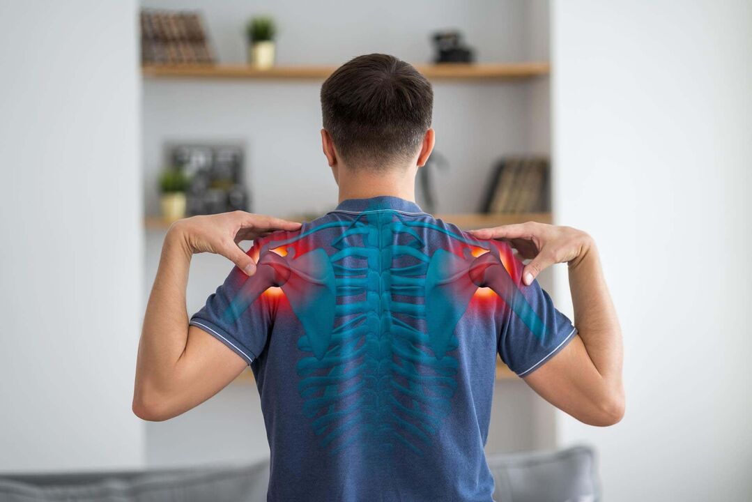
Symptoms of shoulder arthritis

The main clinical symptoms of the shoulder joint are pain, joint stiffness or even complete loss of mobility and joint deformation.The distinctive features of deformed joint pain are:
  • Occurs at the onset of flexion, extension, or rotation;
  • Increased during physical activity;
  • Nocturnal character due to stasis of intraosseous venous blood;
  • Presence of obstruction - sudden jamming of the joint due to separation of fragments of bone and cartilage separated between the joint surfaces;
  • Weather Dependence - Pain that worsens when the weather changes (pain becomes more severe in humid and cold climates).
Arthropathy is a chronic pathology. In the early stages of the disease, pain occurs periodically (as the disease worsens). The rate of pathological progression depends on the promptness of treatment initiation and the adequacy of lifestyle changes. Shoulder pain becomes chronic if it persists for 6 months or longer. The change from acute to chronic pain predicts the progression of the pathological process.

Causes of Shoulder Arthritis

Causes of shoulder arthritis fall into two categories:
  1. Modifiable - can be corrected;
  2. Unmodifiable - it is impossible to influence their behavior.
Non-modifiable factors that may increase the risk of joint changes in the shoulder joint include:
  • Gender - before 50 years of age, women are less susceptible to this disease than men; after about 50 years, the prevalence of the pathology in representatives of both sexes is approximately the same;
  • Age of the person - the older the patient, the higher the risk (the degeneration process of cartilage tissue proceeds faster than the regeneration process starting around 30 years old, which creates prerequisites for the development of the disease);
  • Congenital anomalies of the shoulder structure - increased mobility (hypermobility), underdevelopment of the connective tissue (usually the articular cartilage is represented by type 2 collagen fibers, with underdevelopment there is a replacement of less durable collagen types), joint instability;
  • Genetic Features - Genetically determined polymorphisms in the type 2 collagen dominance, interleukin 1 and interleukin 2 genes.
Modifiable risk factors for right or left shoulder joint deformity include:
  • Traumatic joint injuries;
  • Excessive physical activity (strength sports and martial arts, including barbell bench press);
  • Obesity - For the shoulder joint, the important factor is not the increase in mechanical load, but the metabolic changes that occur in the connective tissue, incl. The chronic inflammatory state that accompanies obesity;
  • Weak shoulder muscles, especially in those who perform precise activities with their hands (jewellers, dentists, secretaries, writers);
  • Lack of vitamin D, which is actively involved in maintaining the health of the musculoskeletal system;
  • A diet low in vitamin C, which is an important part of the body’s calcium and phosphorus metabolism;
  • Hormonal imbalances – thyroid disease, diabetes, etc. ;
  • Smoking – active and passive smoking.
In the shoulder joint, the main targets of pathological processes are articular cartilage, subchondral bone, and synovium. In affected cartilage, the synthesis of proteoglycans is reduced, fragmentation and rupture of the plates are observed, exposing the underlying bone. Increased non-physiological loading on the bones can lead to bone compaction, the development of cysts and osteophytes (marginal growth).

diagnosis

Patients with shoulder pain should start with an X-ray. It is important to scan multiple projections to examine the joint in detail. Images can be taken in direct projection, internally rotated and externally rotated positions. To assess the soft tissue structure of a joint, especially in the early stages of arthrosis, an ultrasound scan of the joint is most informative. If the diagnosis remains unclear, joint magnetic resonance imaging/computed tomography is recommended. In the next stage, preservation of joint function is assessed.

Expert Opinion

All morphogenesis of joints is involved in pathological processes. The main symptom of osteoarthritis is pain in the joint area, caused not only by synovitis, but also by bone damage (osteitis, periostitis), involvement of the soft tissues around the joint (tendonitis, tenosynovitis, myalgia, entheseal lesions, joint capsule tension). Stretch) caused. , meniscal degeneration, and neurosensory system involvement (e. g. , large osteophytes irritating nerve trunks). Therefore, the sooner treatment begins, incl. By making lifestyle changes, you can more effectively control pain.

treat

In the initial stages of the pathological process, joint treatment of the shoulder joint is performed conservatively, and with severe degeneration of the articular cartilage, surgical intervention (endoprosthesis) is required.

Conservative treatment

During an exacerbation, the first priority is pain relief. NSAIDs are most commonly used to relieve pain. They can be used topically (in the form of creams and ointments), injected into the joint space, or used systemically (tablets, intramuscular injection). In some patients, the pain may be so severe that a short course of corticosteroid medication may be used to relieve the pain.Intra-articular injections of hyaluronic acid or plasma, including. Rich in platelets, it can have a stimulating effect on the cartilage plate and promote its renewal (this treatment is considered pathogenic). These injections help speed up the synthesis of collagen and elastin fibers that form the basis of cartilage. As a result, the structure of the cartilage layer and synovium is improved, which helps to increase the consistency of the joint surface. These intra-articular injections help optimize the production of synovial fluid, which not only absorbs shock and moisturizes cartilage, but also improves the metabolic processes of chondrocytes and increases their internal potential. After the acute process has subsided, physiotherapy rehabilitation methods (pulsed current, ultrasound and laser therapy) can be used as part of complex treatment. These surgeries have complex positive effects on joint structures.

Surgery

The surgery causes severe damage to the cartilage plates and is associated with ongoing pain and joint dysfunction, resulting in the inability to care for oneself and perform professional tasks. The modern approach to surgical intervention in the shoulder joint is the placement of an endoprosthesis. At SM-Clinic, surgeries are performed strictly following the use of the latest generation of endoprostheses. This is key to achieving optimal treatment results.

Prevent shoulder joint arthritis

Primary prevention of shoulder arthritis aims to maintain optimal metabolism of the osteochondral compartment. For this purpose it is recommended:
  • Maintain a normal weight;
  • Fully compensate for endocrine disorders in the body (endocrinologist consultation and dynamic monitoring are required);
  • Dose-strengthening muscle girdle of shoulder girdle;
  • If your professional activities involve performing similar movements on your shoulders, warm up regularly.
To prevent the progression of shoulder arthrosis, the following recommendations are important:
  • Avoid lifting heavy objects, including. barbell push-ups;
  • Undertake repeated therapeutic massage sessions;
  • Participate in regular health-promoting gymnastics (under the supervision of a physical therapy specialist).

recovery

After the prosthesis is placed, a plaster cast is applied to provide the necessary degree of fixation. After the cast is removed, the recovery period for joint functional activities begins. For this purpose, it is recommended to carry out therapeutic massage, physiotherapy and health gymnastics courses under the supervision of a physiotherapist.

Questions and Answers

Which doctor treats shoulder arthritis?

The disease is diagnosed and treated by an orthopedic traumatologist.

Representatives of which professions most often suffer from shoulder joint disease?

Athletes who engage in sports such as volleyball, tennis, basketball, throwing, and reloading are at greatest risk for degenerative dystrophic destruction of the cartilage layer of the shoulder joint.

Does shoulder pain signal the development of arthropathy?

In fact, pain is the main symptom of joint disease. However, pain can also be a symptom of other conditions—adhesive capsulitis, osteoarthritis, rotator cuff muscle injury, etc. A qualified orthopedic traumatologist will help you make the correct diagnosis and choose treatment.